
Ferroptosis-centered Drug Response Information
General Information of the Drug (ID: ferrodrug0226)
| Name |
Dihydroartemisinin
|
||||
|---|---|---|---|---|---|
| Synonyms |
Dihydroartemisinin; 71939-50-9; Artenimol; Cotexin; Dihydroqinghaosu; Alaxin; Cotecxin; Dihydroartemisinine; 81496-82-4; Dynamax; Salaxin; Santecxin; (3R,5aS,6R,8aS,9R,10S,12R,12aR)-3,6,9-trimethyldecahydro-3H-3,12-epoxy[1,2]dioxepino[4,3-i]isochromen-10-ol; Di-hydroqinghaosu; GNF-PF-5634; Artenimol [INN]; Dihydro Artemisinin; (3r,5as,6r,8as,9r,10s,12r,12ar)-decahydro-3,6,9-trimethyl-3,12-epoxy-12h-pyrano[4,3-j][1,2]benzodioxepin-10-ol; .beta.-dihydroartemisinin; Dihydroartemisinin, .beta.-; 6A9O50735X; 3,12-Epoxy-12H-pyrano(4,3-j)-1,2-benzodioxepin-10-ol, decahydro-3,6,9-trimethyl-, (3R,5aS,6R,8aS,9R,10S,12R,12aR)-; 3,12-Epoxy-12H-pyrano[4,3-j]-1,2-benzodioxepin-10-ol, decahydro-3,6,9-trimethyl-, (3R,5aS,6R,8aS,9R,10S,12R,12aR)-; b-Dihydroartemisinin; ARTENIMOL [MART.]; ARTENIMOL [WHO-DD]; ARTENIMOL [WHO-IP]; DIHYDROARTEMISININ [MI]; GTPL9957; SCHEMBL17156483; CHEBI:135921; DTXSID501021652; ARTENIMOLUM [WHO-IP LATIN]; DIHYDROARTEMISININ [USP-RS]; DIHYDROARTEMISININ [WHO-DD]; HY-N0176; AKOS032949625; CS-5595; FT-0649320; EURARTESIM COMPONENT DIHYDROARTEMISININ; EN300-7419972; BRD-K62213621-001-01-6; (1R,4S,5R,8S,9R,10S,12R,13R)-1,5,9-trimethyl-11,14,15,16-tetraoxatetracyclo[10.3.1.0^{4,13}.0^{8,13}]hexadecan-10-ol; (3R,5aS,6R,8aS,9R,10S,12R,12aR)-3,6,9-trimethyldecahydro-12H-3,12-epoxy[1,2]dioxepino[4,3-i]isochromen-10-ol; (3R,5AS,6R,8AS,9R,10S,12R,12AR)-3,6,9-TRIMETHYLDECAHYDRO-3,12-EPOXY-12H-PYRANO(4,3-J)-1,2-BENZODIOXEPIN-10-OL
Click to Show/Hide
|
||||
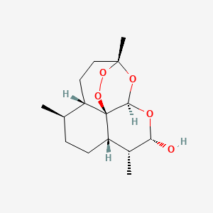
| Structure |
 |
||||
| Formula |
C15H24O5
|
||||
| IUPAC Name |
(1R,4S,5R,8S,9R,10S,12R,13R)-1,5,9-trimethyl-11,14,15,16-tetraoxatetracyclo[10.3.1.04,13.08,13]hexadecan-10-ol
|
||||
| Canonical SMILES |
CC1CCC2C(C(OC3C24C1CCC(O3)(OO4)C)O)C
|
||||
| InChI |
InChI=1S/C15H24O5/c1-8-4-5-11-9(2)12(16)17-13-15(11)10(8)6-7-14(3,18-13)19-20-15/h8-13,16H,4-7H2,1-3H3/t8-,9-,10+,11+,12+,13-,14-,15-/m1/s1
|
||||
| InChIKey |
BJDCWCLMFKKGEE-ISOSDAIHSA-N
|
||||
| PubChem CID | |||||
Full List of Ferroptosis Target Related to This Drug
Phospholipid hydroperoxide glutathione peroxidase (GPX4)
| In total 7 item(s) under this Target | |||||
| Experiment 1 Reporting the Ferroptosis-centered Drug Act on This Target | [1] | ||||
| Target for Ferroptosis | Marker/Suppressor | ||||
| Responsed Disease | Glioblastoma | ICD-11: 2A00 | |||
| Responsed Regulator | Endoplasmic reticulum chaperone BiP (HSPA5) | Suppressor | |||
| Pathway Response | Fatty acid metabolism | hsa01212 | |||
| Ferroptosis | hsa04216 | ||||
| Apoptosis | hsa04210 | ||||
| Cell Process | Cell ferroptosis | ||||
| Cell apoptosis | |||||
| Cell proliferation | |||||
| In Vitro Model | U-251MG cells | Astrocytoma | Homo sapiens | CVCL_0021 | |
| U-373MG cells | Astrocytoma | Homo sapiens | CVCL_2219 | ||
| HT22 cells | Normal | Mus musculus | CVCL_0321 | ||
| In Vivo Model |
Specific pathogen-free athymic nude BALB/c mice (4-6 weeks old) were obtained from Guangdong Experimental Animal Centre (Guangzhou, China). To generate murine subcutaneous tumors, cells (for U251: 2 x 10⁶ cells; for U373: 2 x 10⁶ cells) were suspended in 0.2 ml PBS and injected into the flanks of mice (n = 6/group). Tumor volume was measured once every 3 days using calipers.
Click to Show/Hide
|
||||
| Response regulation | HSPA5 upregulation increased the expression and activity of glutathione peroxidase 4 (GPX4), which neutralized Dihydroartemisinin-induced lipid peroxidation and thus protected glioma cells from ferroptosis. Ferroptosis might be a novel anticancer mechanism of DHA in glioma and HSPA5 may serve as a negative regulator of DHA-induced ferroptosis. | ||||
| Experiment 2 Reporting the Ferroptosis-centered Drug Act on This Target | [1] | ||||
| Target for Ferroptosis | Marker/Suppressor | ||||
| Responsed Disease | Glioblastoma | ICD-11: 2A00 | |||
| Responsed Regulator | Endoplasmic reticulum chaperone BiP (HSPA5) | Suppressor | |||
| Pathway Response | Fatty acid metabolism | hsa01212 | |||
| Ferroptosis | hsa04216 | ||||
| Apoptosis | hsa04210 | ||||
| Cell Process | Cell ferroptosis | ||||
| Cell apoptosis | |||||
| Cell proliferation | |||||
| In Vitro Model | U-251MG cells | Astrocytoma | Homo sapiens | CVCL_0021 | |
| U-373MG cells | Astrocytoma | Homo sapiens | CVCL_2219 | ||
| HT22 cells | Normal | Mus musculus | CVCL_0321 | ||
| In Vivo Model |
Specific pathogen-free athymic nude BALB/c mice (4-6 weeks old) were obtained from Guangdong Experimental Animal Centre (Guangzhou, China). To generate murine subcutaneous tumors, cells (for U251: 2 x 10⁶ cells; for U373: 2 x 10⁶ cells) were suspended in 0.2 ml PBS and injected into the flanks of mice (n = 6/group). Tumor volume was measured once every 3 days using calipers.
Click to Show/Hide
|
||||
| Response regulation | HSPA5 upregulation increased the expression and activity of glutathione peroxidase 4 (GPX4), which neutralized Dihydroartemisinin-induced lipid peroxidation and thus protected glioma cells from ferroptosis. Ferroptosis might be a novel anticancer mechanism of DHA in glioma and HSPA5 may serve as a negative regulator of DHA-induced ferroptosis. | ||||
| Experiment 3 Reporting the Ferroptosis-centered Drug Act on This Target | [6] | ||||
| Target for Ferroptosis | Suppressor | ||||
| Responsed Disease | Glioblastoma | ICD-11: 2A00 | |||
| Pathway Response | Fatty acid metabolism | hsa01212 | |||
| Ferroptosis | hsa04216 | ||||
| Cell Process | Cell ferroptosis | ||||
| Cell proliferation | |||||
| In Vitro Model | U87 MG-Red-Fluc cells | Glioblastoma | Homo sapiens | CVCL_5J12 | |
| A-172 cells | Glioblastoma | Homo sapiens | CVCL_0131 | ||
| Response regulation | Dihydroartemisinin (DHA) had a selective killing effect on glioblastoma, which was associated with over-expression of transferrin receptors. The primary mechanism by which DHA caused ferroptosis was down-regulation of GPX4 and the following lipid ROS accumulation. | ||||
| Experiment 4 Reporting the Ferroptosis-centered Drug Act on This Target | [2] | ||||
| Target for Ferroptosis | Suppressor | ||||
| Responsed Disease | Glioblastoma | ICD-11: 2A00 | |||
| Pathway Response | Ferroptosis | hsa04216 | |||
| Cell Process | Cell ferroptosis | ||||
| In Vitro Model | U87 MG-Red-Fluc cells | Glioblastoma | Homo sapiens | CVCL_5J12 | |
| U-251MG cells | Astrocytoma | Homo sapiens | CVCL_0021 | ||
| In Vivo Model |
All BALB/C nude mice were purchased from Huafukang Biotechnology (Beijing, China). These mice were 5 weeks old and weighed 14-16 g. We established subcutaneous tumour-forming mouse model by injecting 5 x 10⁶ U87 cells into the lateral abdomen of BALB/C nude mice. Animals were then treated with DHA solvent (50 mg/kg) by intragastric administration once a day for 26 days.
Click to Show/Hide
|
||||
| Response regulation | Dihydroartemisinin could promote ferroptosis in glioma cells. Low expression of GPX4 and high expression of HMOX1 were identified in DHA treated glioma cells. MAZ was further identified as the direct target of long noncoding RNA (lncRNA) TUG1 through luciferase assay. Downregulated expression of TUG1 and upregulated expression of MAZ were identified in DHA treated glioma cells. TUG1 overexpression or inhibition of FTH1 expression could enhance the antiglioma effect of DHA in vitro and in vivo, providing a promising strategy to enhance the antitumor effect of DHA in glioma. | ||||
| Experiment 5 Reporting the Ferroptosis-centered Drug Act on This Target | [7] | ||||
| Target for Ferroptosis | Suppressor | ||||
| Responsed Disease | Lung cancer | ICD-11: 2C25 | |||
| Pathway Response | Fatty acid metabolism | hsa01212 | |||
| Ferroptosis | hsa04216 | ||||
| Autophagy | hsa04140 | ||||
| Cell Process | Cell ferroptosis | ||||
| Cell proliferation | |||||
| Cell autophagy | |||||
| In Vitro Model | NCI-H292 cells | Lung mucoepidermoid carcinoma | Homo sapiens | CVCL_0455 | |
| HCT 116 cells | Colon carcinoma | Homo sapiens | CVCL_0291 | ||
| HT29 cells | Colon cancer | Mus musculus | CVCL_A8EZ | ||
| SW480 cells | Colon adenocarcinoma | Homo sapiens | CVCL_0546 | ||
| MDA-MB-453 cells | Breast adenocarcinoma | Homo sapiens | CVCL_0418 | ||
| MCF-7 cells | Breast carcinoma | Homo sapiens | CVCL_0031 | ||
| HT-1080 cells | Fibrosarcoma | Homo sapiens | CVCL_0317 | ||
| In Vivo Model |
GPX4 iKO H292 cells were inoculated by injecting 3 x 10⁶ cells in 0.1 mL PBS subcutaneously in the right flank of six- to eight-week-old female athymic nude Foxn1nu/Foxn1 mice (Envigo, East Millstone, NJ, USA). Following inoculation, the mice were monitored until they have fully recovered and are moving. Mice were randomly allocated into their respective groups (non-blinded). Tumor growth was monitored regularly via external caliper measurements.
Click to Show/Hide
|
||||
| Response regulation | Dihydroartemisinin (DAT) can augment GPX4 inhibition-induced ferroptosis in a cohort of cancer cells that are otherwise highly resistant to ferroptosis. Collectively, artemisinin compounds can sensitize cells to ferroptosis by regulating cellular iron homeostasis in Lung mucoepidermoid carcinoma. | ||||
| Experiment 6 Reporting the Ferroptosis-centered Drug Act on This Target | [8] | ||||
| Target for Ferroptosis | Suppressor | ||||
| Responsed Disease | Lung cancer | ICD-11: 2C25 | |||
| Pathway Response | Ferroptosis | hsa04216 | |||
| Cell Process | Cell ferroptosis | ||||
| Cell apoptosis | |||||
| Cell proliferation | |||||
| In Vitro Model | A-549 cells | Lung adenocarcinoma | Homo sapiens | CVCL_0023 | |
| HCC827 cells | Lung adenocarcinoma | Homo sapiens | CVCL_2063 | ||
| NCI-H1975 cells | Lung adenocarcinoma | Homo sapiens | CVCL_1511 | ||
| Response regulation | Dihydroartemisinin (DHA) treatment decreased the levels of GPX4, DHA significantly induced apoptosis and ferroptosis in a dose-dependent manner and exhibited high cellular toxicity on A549-GR (non-small cell lung cancer) cells when combined with gefitinib. | ||||
| Experiment 7 Reporting the Ferroptosis-centered Drug Act on This Target | [9] | ||||
| Target for Ferroptosis | Suppressor | ||||
| Responsed Disease | Cervical cancer | ICD-11: 2C77 | |||
| Pathway Response | Fatty acid metabolism | hsa01212 | |||
| Ferroptosis | hsa04216 | ||||
| Cell Process | Cell ferroptosis | ||||
| In Vitro Model | HeLa cells | Endocervical adenocarcinoma | Homo sapiens | CVCL_0030 | |
| SiHa cells | Cervical squamous cell carcinoma | Homo sapiens | CVCL_0032 | ||
| Response regulation | Dihydroartemisinin (DHA) treatment initiated ferroptosis, as evidenced by the accumulation of reactive oxygen species (ROS), malondialdehyde (MDA) and liquid peroxidation (LPO) levels and simultaneously depletion of glutathione peroxidase 4 (GPX4) and glutathione (GSH). Moreover, nuclear receptor coactivator 4 (NCOA4)-mediated ferritinophagy was also induced by DHA leading to subsequent increases of intracellular labile iron pool (LIP), exacerbated the Fenton reaction resulting in excessive ROS production, and enhanced cervical cancer ferroptosis. | ||||
Ferritin heavy chain (FTH1)
| In total 2 item(s) under this Target | |||||
| Experiment 1 Reporting the Ferroptosis-centered Drug Act on This Target | [2] | ||||
| Target for Ferroptosis | Marker | ||||
| Responsed Disease | Glioblastoma | ICD-11: 2A00 | |||
| Responsed Regulator | Myc-associated zinc finger protein (MAZ) | Driver | |||
| Pathway Response | Ferroptosis | hsa04216 | |||
| Cell Process | Cell ferroptosis | ||||
| In Vitro Model | U87 MG-Red-Fluc cells | Glioblastoma | Homo sapiens | CVCL_5J12 | |
| U-251MG cells | Astrocytoma | Homo sapiens | CVCL_0021 | ||
| In Vivo Model |
All BALB/C nude mice were purchased from Huafukang Biotechnology (Beijing, China). These mice were 5 weeks old and weighed 14-16 g. We established subcutaneous tumour-forming mouse model by injecting 5 x 10⁶ U87 cells into the lateral abdomen of BALB/C nude mice. Animals were then treated with DHA solvent (50 mg/kg) by intragastric administration once a day for 26 days.
Click to Show/Hide
|
||||
| Response regulation | Dihydroartemisinin (DHA) could promote ferroptosis in glioma cells. Low expression of GPX4 and high expression of HMOX1 were identified in DHA treated glioma cells. MAZ was further identified as the direct target of long noncoding RNA (lncRNA) TUG1 through luciferase assay. Downregulated expression of TUG1 and upregulated expression of MAZ were identified in DHA treated glioma cells. TUG1 overexpression or inhibition of FTH1 expression could enhance the antiglioma effect of DHA in vitro and in vivo, providing a promising strategy to enhance the antitumor effect of DHA in glioma. | ||||
| Experiment 2 Reporting the Ferroptosis-centered Drug Act on This Target | [2] | ||||
| Target for Ferroptosis | Marker | ||||
| Responsed Disease | Glioblastoma | ICD-11: 2A00 | |||
| Responsed Regulator | TUG1 (IncRNA) | Suppressor | |||
| Pathway Response | Ferroptosis | hsa04216 | |||
| Cell Process | Cell ferroptosis | ||||
| In Vitro Model | U87 MG-Red-Fluc cells | Glioblastoma | Homo sapiens | CVCL_5J12 | |
| U-251MG cells | Astrocytoma | Homo sapiens | CVCL_0021 | ||
| In Vivo Model |
All BALB/C nude mice were purchased from Huafukang Biotechnology (Beijing, China). These mice were 5 weeks old and weighed 14-16 g. We established subcutaneous tumour-forming mouse model by injecting 5 x 10⁶ U87 cells into the lateral abdomen of BALB/C nude mice. Animals were then treated with DHA solvent (50 mg/kg) by intragastric administration once a day for 26 days.
Click to Show/Hide
|
||||
| Response regulation | Dihydroartemisinin (DHA) could promote ferroptosis in glioma cells. Low expression of GPX4 and high expression of HMOX1 were identified in DHA treated glioma cells. MAZ was further identified as the direct target of long noncoding RNA (lncRNA) TUG1 through luciferase assay. Downregulated expression of TUG1 and upregulated expression of MAZ were identified in DHA treated glioma cells. TUG1 overexpression or inhibition of FTH1 expression could enhance the antiglioma effect of DHA in vitro and in vivo, providing a promising strategy to enhance the antitumor effect of DHA in glioma. | ||||
Unspecific Target
| In total 3 item(s) under this Target | |||||
| Experiment 1 Reporting the Ferroptosis-centered Drug Act on This Target | [3] | ||||
| Responsed Disease | Acute myeloid leukaemia | ICD-11: 2A60 | |||
| Responsed Regulator | 5'-AMP-activated protein kinase catalytic subunit alpha-1 (PRKAA1) | Driver | |||
| Pathway Response | Fatty acid metabolism | hsa01212 | |||
| Ferroptosis | hsa04216 | ||||
| Autophagy | hsa04140 | ||||
| Cell Process | Cell ferroptosis | ||||
| Cell autophagy | |||||
| Cell proliferation | |||||
| Cell cycle | |||||
| In Vitro Model | HL-60 cells | Adult acute myeloid leukemia | Homo sapiens | CVCL_0002 | |
| KG1 cells | Normal | Mus musculus | CVCL_UD72 | ||
| THP-1 cells | Childhood acute monocytic leukemia | Homo sapiens | CVCL_0006 | ||
| In Vivo Model |
BALB/c Nude Mice (4 weeks old) were obtained from Shanghai Experimental Animal Center of the Chinese Academy of Sciences (Shanghai, China) and then subcutaneously injection with HL60 cells (1 x 10⁷, suspended in 0.1 mL PBS). After tumors reached 100-200 mm3, the mice were randomly assigned to two groups. DHA was administered intraperitoneal injection once a day at 50 mg/kg body weight and the mice in normal control were received equal amounts of vehicle (10% DMSO in sterile corn oil). On the 28th day, mice were euthanized. The tumor volumes were measured every 4 days with a caliper.
Click to Show/Hide
|
||||
| Response regulation | Dihydroartemisinin (DHA) strongly inhibited the viability of acute myeloid leukemia (AML) cell lines and arrest cell cycle at G0/G1 phase. Mechanistically, DHA induced autophagy by regulating the activity of AMPK/mTOR/p70S6k signaling pathway, which accelerated the degradation of ferritin, increased the labile iron pool, promoted the accumulation of cellular ROS and eventually led to ferroptotic cell death. | ||||
| Experiment 2 Reporting the Ferroptosis-centered Drug Act on This Target | [4] | ||||
| Responsed Disease | Acute myeloid leukaemia | ICD-11: 2A60 | |||
| Responsed Regulator | Metallothionein-1A (MT1A) | . | |||
| Pathway Response | Ferroptosis | hsa04216 | |||
| Cell Process | Cell ferroptosis | ||||
| In Vitro Model | MOLM-14 cells | Leukemia | Homo sapiens | CVCL_7916 | |
| OCI-AML-2 cells | Acute myeloid leukemia | Homo sapiens | CVCL_1619 | ||
| HL-60 cells | Adult acute myeloid leukemia | Homo sapiens | CVCL_0002 | ||
| SET-2 cells | Acute megakaryoblastic leukemia | Homo sapiens | CVCL_2187 | ||
| MV4-11 cells | Childhood acute monocytic leukemia | Homo sapiens | CVCL_0064 | ||
| K-562 cells | Chronic myelogenous leukemia | Homo sapiens | CVCL_0004 | ||
| THP-1 cells | Childhood acute monocytic leukemia | Homo sapiens | CVCL_0006 | ||
| UT-7/Epo cells | Acute megakaryoblastic leukemia | Homo sapiens | CVCL_5202 | ||
| SKM-1 cells | Acute myeloid leukemia | Homo sapiens | CVCL_0098 | ||
| NB4 cells | Acute promyelocytic leukemia | Homo sapiens | CVCL_0005 | ||
| Kasumi-1 cells | Acute myeloid leukemia | Homo sapiens | CVCL_0589 | ||
| Response regulation | Dihydroartemisinin (DHA) activated zinc metabolism signaling, especially the upregulation of metallothionein (MT). DHA activates ferritinophagy and subsequent ferroptosis in AML and that MTs are involved in glutathione regenerating and antioxidant response in Acute Myeloid Leukemia. | ||||
| Experiment 3 Reporting the Ferroptosis-centered Drug Act on This Target | [5] | ||||
| Responsed Disease | Pancreatic cancer | ICD-11: 2C10 | |||
| Responsed Regulator | Cellular tumor antigen p53 (TP53) | Driver | |||
| Pathway Response | Fatty acid metabolism | hsa01212 | |||
| Cell Process | Cell ferroptosis | ||||
| Cell proliferation | |||||
| In Vitro Model | Panc02 cells | Pancreatic ductal adenocarcinoma | Mus musculus | CVCL_D627 | |
| PANC-1 cells | Pancreatic ductal adenocarcinoma | Homo sapiens | CVCL_0480 | ||
| In Vivo Model |
Six to eight-week-old female C57BL/6 mice were purchased from the Experimental Animal Center of Military Medical Sciences (Beijing, China). C57BL/6 mice were anesthetized and the tail of the pancreas was exposed. Panc 02 cells were resuspended in PBS at a concentration of 1 x 10⁶ cells/0.1 ml and 50 ul cells were injected into the tail of the pancreas. Tumor-bearing mice were randomly divided into two groups (3 days after implantation). The control group was intraperitoneally injected 200 ul PBS daily for 10 days, and the DHA group was intraperitoneally injected with 100 mg/kg DHA daily for 10 days. The pancreatic tumors and spleens of the mice were collected for subsequent analysis.
Click to Show/Hide
|
||||
| Response regulation | Dihydroartemisinin has anti-tumor effect in pancreatic cancer cells in vitro and in vivo. DHA treatment induced ferroptosis by increasing P53 and AOLX12 expression. | ||||
Polyunsaturated fatty acid lipoxygenase ALOX12 (ALOX12)
| In total 1 item(s) under this Target | |||||
| Experiment 1 Reporting the Ferroptosis-centered Drug Act on This Target | [5] | ||||
| Target for Ferroptosis | Driver | ||||
| Responsed Disease | Pancreatic cancer | ICD-11: 2C10 | |||
| Pathway Response | Fatty acid metabolism | hsa01212 | |||
| Cell Process | Cell ferroptosis | ||||
| Cell proliferation | |||||
| In Vitro Model | Panc02 cells | Pancreatic ductal adenocarcinoma | Mus musculus | CVCL_D627 | |
| PANC-1 cells | Pancreatic ductal adenocarcinoma | Homo sapiens | CVCL_0480 | ||
| In Vivo Model |
Six to eight-week-old female C57BL/6 mice were purchased from the Experimental Animal Center of Military Medical Sciences (Beijing, China). C57BL/6 mice were anesthetized and the tail of the pancreas was exposed. Panc 02 cells were resuspended in PBS at a concentration of 1 x 10⁶ cells/0.1 ml and 50 ul cells were injected into the tail of the pancreas. Tumor-bearing mice were randomly divided into two groups (3 days after implantation). The control group was intraperitoneally injected 200 ul PBS daily for 10 days, and the DHA group was intraperitoneally injected with 100 mg/kg DHA daily for 10 days. The pancreatic tumors and spleens of the mice were collected for subsequent analysis.
Click to Show/Hide
|
||||
| Response regulation | Dihydroartemisinin has anti-tumor effect in pancreatic cancer cells in vitro and in vivo. DHA treatment induced ferroptosis by increasing P53 and AOLX12 expression. | ||||
Nuclear receptor coactivator 4 (NCOA4)
| In total 1 item(s) under this Target | ||||
| Experiment 1 Reporting the Ferroptosis-centered Drug Act on This Target | [9] | |||
| Target for Ferroptosis | Driver | |||
| Responsed Disease | Cervical cancer | ICD-11: 2C77 | ||
| Pathway Response | Fatty acid metabolism | hsa01212 | ||
| Ferroptosis | hsa04216 | |||
| Cell Process | Cell ferroptosis | |||
| In Vitro Model | HeLa cells | Endocervical adenocarcinoma | Homo sapiens | CVCL_0030 |
| SiHa cells | Cervical squamous cell carcinoma | Homo sapiens | CVCL_0032 | |
| Response regulation | Dihydroartemisinin (DHA) treatment initiated ferroptosis, as evidenced by the accumulation of reactive oxygen species (ROS), malondialdehyde (MDA) and liquid peroxidation (LPO) levels and simultaneously depletion of glutathione peroxidase 4 (GPX4) and glutathione (GSH). Moreover, nuclear receptor coactivator 4 (NCOA4)-mediated ferritinophagy was also induced by DHA leading to subsequent increases of intracellular labile iron pool (LIP), exacerbated the Fenton reaction resulting in excessive ROS production, and enhanced cervical cancer ferroptosis. | |||
Heme oxygenase 1 (HMOX1)
| In total 1 item(s) under this Target | |||||
| Experiment 1 Reporting the Ferroptosis-centered Drug Act on This Target | [2] | ||||
| Target for Ferroptosis | Driver | ||||
| Responsed Disease | Glioblastoma | ICD-11: 2A00 | |||
| Pathway Response | Ferroptosis | hsa04216 | |||
| Cell Process | Cell ferroptosis | ||||
| In Vitro Model | U87 MG-Red-Fluc cells | Glioblastoma | Homo sapiens | CVCL_5J12 | |
| U-251MG cells | Astrocytoma | Homo sapiens | CVCL_0021 | ||
| In Vivo Model |
All BALB/C nude mice were purchased from Huafukang Biotechnology (Beijing, China). These mice were 5 weeks old and weighed 14-16 g. We established subcutaneous tumour-forming mouse model by injecting 5 x 10⁶ U87 cells into the lateral abdomen of BALB/C nude mice. Animals were then treated with DHA solvent (50 mg/kg) by intragastric administration once a day for 26 days.
Click to Show/Hide
|
||||
| Response regulation | Dihydroartemisinin could promote ferroptosis in glioma cells. Low expression of GPX4 and high expression of HMOX1 were identified in DHA treated glioma cells. MAZ was further identified as the direct target of long noncoding RNA (lncRNA) TUG1 through luciferase assay. Downregulated expression of TUG1 and upregulated expression of MAZ were identified in DHA treated glioma cells. TUG1 overexpression or inhibition of FTH1 expression could enhance the antiglioma effect of DHA in vitro and in vivo, providing a promising strategy to enhance the antitumor effect of DHA in glioma. | ||||
Glutathione-specific gamma-glutamylcyclotransferase 1 (CHAC1)
| In total 1 item(s) under this Target | |||||
| Experiment 1 Reporting the Ferroptosis-centered Drug Act on This Target | [10] | ||||
| Target for Ferroptosis | Marker/Driver | ||||
| Responsed Disease | Hepatocellular carcinoma | ICD-11: 2C12 | |||
| Pathway Response | Ferroptosis | hsa04216 | |||
| Cell Process | Cell ferroptosis | ||||
| Cell proliferation | |||||
| In Vitro Model | Hep 3B2.1-7 cells | Hepatocellular carcinoma | Homo sapiens | CVCL_0326 | |
| Huh-7 cells | Hepatocellular carcinoma | Homo sapiens | CVCL_0336 | ||
| Hep-G2 cells | Hepatoblastoma | Homo sapiens | CVCL_0027 | ||
| PLC/PRF/5 cells | Hepatocellular carcinoma | Homo sapiens | CVCL_0485 | ||
| PLC/PRF/5 cells | Hepatocellular carcinoma | Homo sapiens | CVCL_0485 | ||
| In Vivo Model |
A total of 32 male BALB/c nude mice (age, 6-8 weeks; weight, 18-20 g) were obtained from HFK Bioscience Co. Ltd. and housed in a specific pathogen-free facility. The mice were randomly divided into four groups (n = 8/group) and PLC cells were subcutaneously injected into the nude mice. When the xenografted tumors had grown to 80-100 mm3, half of the mice in each group were administered with 100 mg/kg DHA for 5 days/week by gavage.
Click to Show/Hide
|
||||
| Response regulation | Dihydroartemisinin triggers ferroptosis in primary liver cancer cells by promoting and unfolded protein responseinduced upregulation of CHAC1 expression. DHA also promoted the transcription of CHAC1. | ||||
References